


Portable Ultrasound Machine: B-Mode vs Color Doppler
- 2026-09-24
- 64
- Guangzhou Sonostar Technologies Co., Limited
"B-mode" and "color Doppler" are often treated as one thing, yet they answer different clinical questions. When evaluating a portable ultrasound machine, the distinction matters: B-mode renders tissue structure in grayscale, while color Doppler adds blood-flow information on top of the same image. It is not an academic point — it decides which portable ultrasound machine fits a clinic's workload. Published evidence offers practical guidance: a pictorial review of pocket-sized versus standard machines found acceptable image quality for gallstones, intrahepatic duct stones, fluid collections and the abdominal aorta, while solid-organ parenchymal assessment remained limited. This article explains how the two modes differ, when B-mode is enough, and what it means for selecting a portable ultrasound machine.
How B-Mode and Color Doppler Work
Both modes start from the same physical basis: ultrasound pulses reflect off tissue interfaces, and the returning echoes build a grayscale image. B-mode displays these echoes as brightness levels, outlining organs, lesions and fluid. Color Doppler goes further by using the Doppler effect — sound frequency shifts as blood cells move — to estimate motion. The result is a color overlay on the grayscale image showing flow direction and velocity, plus spectral (PW) Doppler for quantitative velocity readings and power Doppler (PDI) for low-flow sensitivity.
The Difference in One Sentence
B-mode answers "what structure is here"; color Doppler answers "is there blood flow, and which way does it move." For a thyroid nodule, B-mode shows the lesion; color Doppler reveals whether it carries internal vascularity. For a gallbladder, B-mode alone usually suffices.
When Is B-Mode Enough?
Certain exams rely on high-contrast echoes where grayscale performs well. The pictorial review cited above reported satisfactory identification of gallstones, intrahepatic duct stones, intra-abdominal fluid, pleural effusion and the abdominal aorta with a pocket-sized B-mode device. Screening scenarios — stones, effusions, gross anatomy — can be served by B-mode alone. The same review noted that parenchymal echotexture and space-occupying lesion assessment were suboptimal compared with standard machines, a limitation worth knowing before purchase.
Why Color Doppler Matters
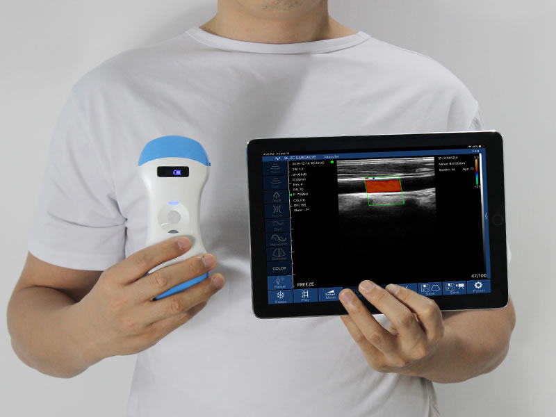
Any exam that depends on flow information requires color Doppler: vascular access assessment, carotid evaluation, thyroid and breast lesion vascularity, and bedside checks where perfusion guides the next step. Randomized trial evidence found a handheld point-of-care device achieved diagnostic accuracy of 89.3% versus 92.5% for a cart-based model, with authors concluding the accuracy of handheld POCUS is similar to that of cart-based systems — but only when the needed mode exists on the machine.
What This Means for a Portable Ultrasound Machine
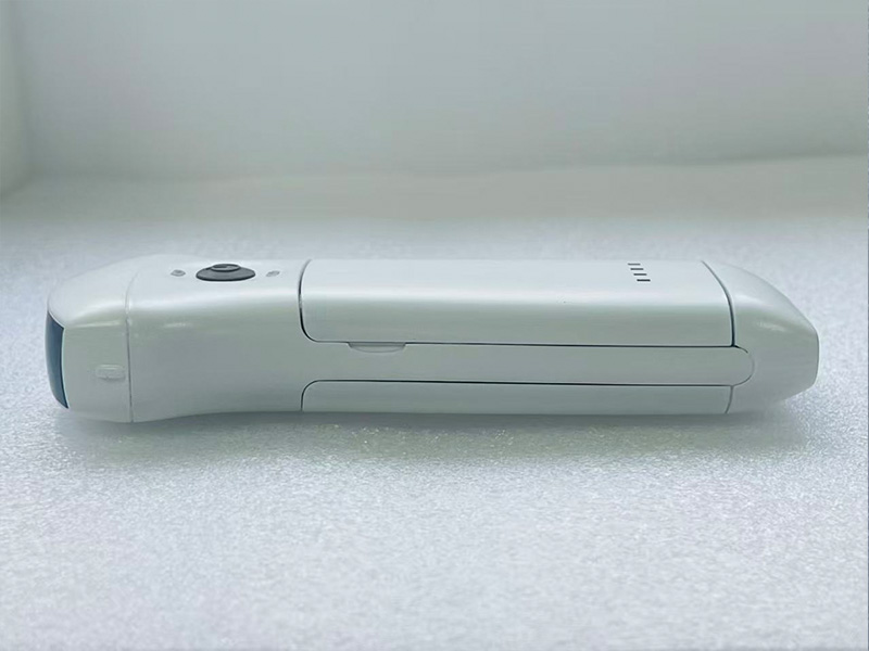
The practical conclusion: instead of choosing between B-mode and color Doppler, most buyers should choose a portable ultrasound machine that offers both. SonoStar wireless probes support B, B/M, Color, PDI and PW display modes in one probe — grayscale screening and flow assessment in the same device. The convex 8C model uses 192 elements and 64 channels at 3.2/5.0 MHz with a 90–305 mm adjustable depth, covering abdominal and general exams; linear models such as the 9N and 9L address thyroid, small parts and superficial vascular work. A single color-capable probe removes the "B-mode or color" trade-off entirely.
Frequently Asked Questions
Is B-mode the same as color Doppler?
No. B-mode displays tissue structure in grayscale; color Doppler adds blood-flow direction and velocity to the same image using the Doppler effect.
Can a portable ultrasound machine detect gallstones?
Yes. Studies found acceptable image quality for gallstones, intrahepatic duct stones and fluid collections with pocket-sized B-mode devices; parenchymal disease assessment is more limited.
Why does my clinic need color Doppler?
Any exam requiring flow information — vascular access, carotid, thyroid or breast lesion vascularity — cannot be performed in B-mode alone.
Do portable ultrasound machines offer both modes?
Most current wireless probes do. SonoStar models support B, B/M, Color, PDI and PW in a single probe, so no trade-off is required.

Choose by Workload, Not by Label
The B-mode versus color Doppler question is really a workload question. Structure-only screening can start with grayscale; anything involving blood flow needs Doppler capability. In the portable segment, machines that combine both modes have become the practical default. SonoStar, founded in 2011 and exporting to more than 100 countries, builds wireless probes with 192-element, 64-channel arrays and B/Color/PDI/PW modes, paired with the SmartUSG app — a reasonable benchmark when evaluating a portable ultrasound machine for your setting.
References
1. Pocket-sized versus standard ultrasound machines in abdominal imaging — pictorial review (Semantic Scholar, GE Vscan vs Philips iU22)
2. Hand-Held Ultrasound Devices Compared with High-End Ultrasound Systems: A Systematic Review — PMC6628329
3. Diagnostic Accuracy of a Handheld Ultrasound vs a Cart-based Model: A Randomized Clinical Trial — PMC11000544
- Pre:Handheld Ultrasound Accuracy: How Reliable Is It Really?
- Next:No more




 Home
Home Product
 News
News Contact