


3D Volume Imaging Wireless Color Ultrasound: Features That Matter and Where It Fits
- 2026-11-10
- 44
- Guangzhou Sonostar Technologies Co., Limited
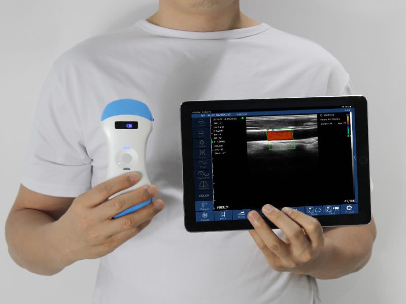
A 3D volume imaging wireless color ultrasound is a probe-shaped system that builds a three-dimensional volume from a convex array and renders it on a phone or tablet, with no trolley, console or separate workstation.
How a probe produces a volume
On a cart-based system the probe emits, a console beamforms, and a workstation renders: three boxes in one room. A 3D volume imaging wireless color ultrasound collapses that chain. Beamforming, battery and radio sit in the scan head, and rendering runs on an iOS tablet or phone connected over the probe's own 802.11g Wi-Fi at 2.4 or 5 GHz.
Acquisition is a sweep. The operator moves the 60° sector across the region of interest, consecutive frames are registered into a volume, and the engine converts voxel intensity into a surface. Volume quality therefore depends on sweep steadiness as much as on electronics. Two display modes are available, B and 3D.
Specifications that matter in practice
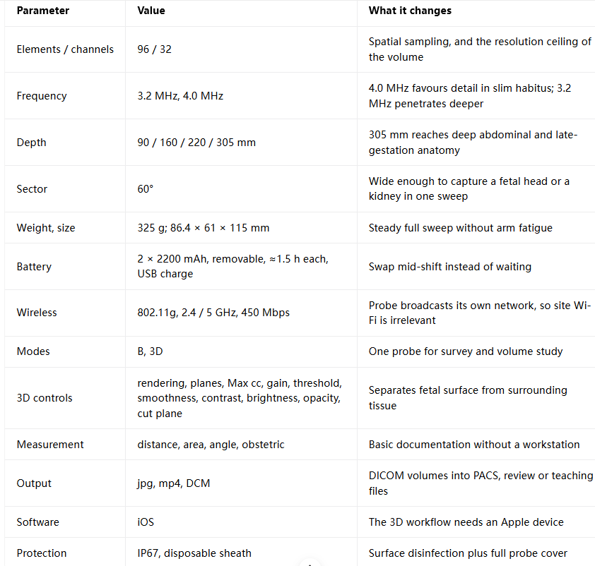
Three of these deserve emphasis. The removable battery means a full sweep does not end when the charge does; the second pack is already in the case. The probe's self-broadcast network matters in field use, emergency vehicles and rural clinics, where there is no Wi-Fi to join. And IP67 with a sheath fits the infection-control routine of a ward rather than the reprocessing cycle of an ultrasound room.

Where a volume earns its place
Obstetrics. Surface rendering shows the fetal face, limbs and position in a form a parent or a referring clinician grasps at once. The axial, sagittal and coronal planes then verify what the surface image hides.
Abdominal organs. Describing a liver, gallbladder, kidney or bladder finding in three orthogonal planes lets a referral note carry position and depth, not just "a mass was seen".
Interventional orientation. Before drainage, biopsy or vascular access, a volume plus multiplanar review helps fix entry point and expected depth. Real-time needle guidance stays two-dimensional.
Primary care and community screening. A clinic with no ultrasound room can still document anatomy with a 3D volume imaging wireless color ultrasound, because the tablet is the display.
Teaching and second opinion. mp4 clips and DCM volumes travel by message; a colleague reviews the same data set rather than a single still frame. The vendor also positions the platform for non-diagnostic keepsake imaging where local rules allow.

FAQ
Is a 3D volume imaging wireless color ultrasound accurate enough for diagnosis?
For volume visualisation and documentation, yes; for primary diagnostic reading, treat it as a triage and communication tool and escalate to a cart system.
Does it need internet access?
No. It uses the Wi-Fi network the probe itself broadcasts.
Can several clinicians view at once?
One device connects at a time; multi-screen sharing depends on platform and firmware.
What does a 3D exam add in time?
A sweep takes seconds; review and rendering take the longer part and can happen away from the bedside.
Guangzhou SonoStar Information Technology Co., Ltd. is a national high-tech enterprise specializing in miniaturized, intelligent ultrasound. We develop and manufacture handheld probe-style intelligent ultrasound imaging diagnostic devices, wearable B-mode ultrasound and other portable systems, supported by long-term industry–academia–research collaboration. CE- and FDA-registered, our products reach more than 100 countries and regions and serve clinical care, primary and emergency medicine, aesthetic medicine and veterinary practice — advancing accessible ultrasound for everyday care. Contact: Ms. Lin, +86 181 2686 2475.
- Pre:3D Volume Imaging Wireless Ultrasound: Features That Matter
- Next:No more




 Home
 Product
 News
 Contact