


3D Volume Imaging Handheld Ultrasound in Primary Care: What the Volume Changes for Frontline Clinici
- 2026-11-12
- 35
- Guangzhou Sonostar Technologies Co., Limited
A 3D volume imaging handheld ultrasound builds a volume from one sweep and renders it on a phone or tablet. In a tertiary hospital that is a convenience; in a township health center or a village clinic it changes what one operator can do, because the constraint at the frontline is not the machine — it is the person holding it. What the volume buys is a second chance to look.
Primary care is the hard case
Three constraints define it. Equipment: a cart is unaffordable and cannot leave the room. Operator: there is no sonographer on site. Distance: the county hospital is an hour away, and the patient may not go.
A 2D handheld probe removes the equipment problem and leaves the operator problem untouched — one slice, at one angle, in real time, decided on the spot. A volume moves part of that decision to after the scan, which is where a small clinic has room to think.
What the volume changes at the frontline
1. Re-slicing replaces on-the-spot judgement. The probe stores the volume, and the operator cuts axial, sagittal and coronal planes afterwards, then asks a senior clinician to review the same dataset. Ambiguous structures get examined without recalling the patient.
2. Obstetric and abdominal screening gains orientation. A rendered fetal profile, limb position or placental location reads in one image and explains to a parent in one sentence; the same view supports family-planning checks and home antenatal visits.
3. Remote support becomes a file transfer. Sending a volume to a county-level reviewer is a different act from describing what you saw; the convex 3D probe stores JPG, MP4 and DICOM files, so the reviewer works from the dataset rather than from the operator's account of it.
4. Supervision works on the same data. Trainees see organ and needle relationships in a rotatable volume; a supervisor marks planes and sends them back — how skill transfers where one experienced clinician works beside three new ones.
What it takes to run one in a village clinic
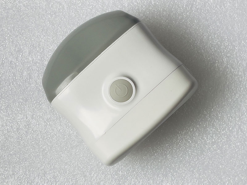
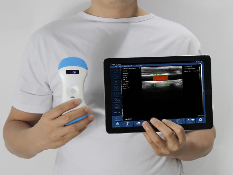
The hardware has to survive the setting, not the showroom. A 325 g convex probe at 86.4 x 61 x 115 mm holds steady through a sweep in one hand. A 60° sector reaching 90 to 305 mm covers basic abdominal and obstetric views. Two 2.200 mAh cells at roughly 1.5 hours each carry a morning. The probe broadcasts its own Wi-Fi, so no clinic network is needed, and an IP67 housing with a single-use sheath keeps cleaning inside routine.
Two limits belong in this paragraph as well: depth and resolution stay below a cart system, and obese patients and deep pelvic views are the known hard cases.
Where 3D still falls short in primary care
The volume does not teach anatomy. An operator who cannot identify a fetal lie on a 2D slice will not identify it in a rendered volume; training remains the binding constraint, and a rollout without it stalls in month two.
It is grayscale. Colour flow sits on the 2D colour models, so a clinic needing vascular assessment should confirm per model that colour Doppler and volume rendering coexist.
It produces no formal report. Images export to a PC for editing, but the device is built for screening and guidance. The referral pathway stays as it was — and that is the local argument for adoption: the county hospital receives earlier, better-described cases, not fewer.
A probe also serves one device at a time, and the 3D app runs on iOS, so the clinic's tablet choice is part of procurement, not an afterthought.

FAQ
Is a 3D volume imaging handheld ultrasound necessary in primary care, or is 2D enough?
2D covers straightforward guidance. The volume earns its place where orientation is the difficulty: fetal position, organ shape, and cases needing a second reader after the scan.
Do operators need sonographer-level certification?
Not for the guidance and screening views described here, provided the clinic has a training plan and a review route. Diagnostic reporting stays with qualified staff.
Does the probe need the clinic network or an internet connection?
No. It runs its own Wi-Fi access point; the network question only arises when images are sent onward.
Can a county hospital review a volume the same day?
Yes, if the clinic exports DICOM and the review workflow is agreed in advance. File size and transfer route are the practical constraints.
Guangzhou SonoStar Information Technology Co., Ltd. is a national high-tech enterprise specializing in miniaturized, intelligent ultrasound. We develop and manufacture handheld probe-style intelligent ultrasound imaging diagnostic devices, wearable B-mode ultrasound and other portable systems, supported by long-term industry–academia–research collaboration. CE- and FDA-registered, our products reach more than 100 countries and regions and serve clinical care, primary and emergency medicine, aesthetic medicine and veterinary practice — advancing accessible ultrasound for everyday care. Contact: Ms. Lin, +86 181 2686 2475.




 Home
 Product
 News
 Contact