


3-in-1 Ultrasound Probe Wireless: One Probe, Three Arrays
- 2026-10-13
- 46
- Guangzhou Sonostar Technologies Co., Limited
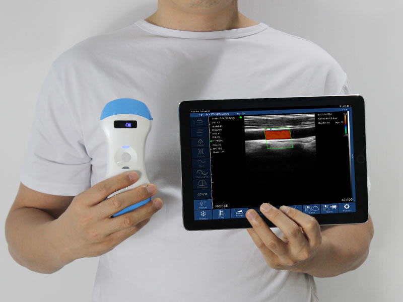
A department that buys handheld ultrasound usually arrives at the same problem quickly: the abdominal exam needs a convex probe, vein cannulation needs a linear probe, and the cardiac window needs a phased array. Buying three probes means three devices to charge, three to disinfect, three to lose, and three lines on the budget. A 3-in-1 ultrasound probe wireless collapses that into one unit — three transducer arrays in a single housing, switching electronically, streaming wirelessly to the phone or tablet already in the clinician's pocket.
What Is a 3-in-1 Ultrasound Probe Wireless?
The "3-in-1" label describes the transducer, not the display. Inside one housing sit three physically distinct arrays:
Convex array — a curved footprint with a wide fan image, for deep abdominal and pelvic structures.
Linear array — a flat, rectangular footprint for superficial, vascular and MSK targets.
Phased array — a small footprint producing a sector (pie-shaped) image, small enough to fit between ribs for cardiac views.
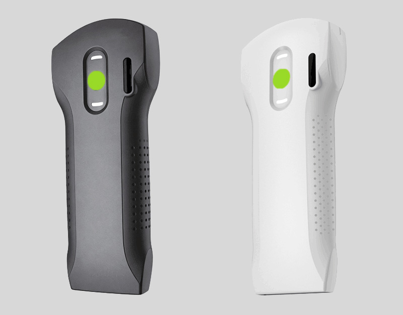
Switching between them is an electronic selection, not a physical swap. On SonoStar's 3-in-1 palm probes, beamforming, image processing, the Wi-Fi transmitter and a rechargeable battery all sit inside the probe body; the image is streamed to an iOS, Android or Windows device over a dual-band Wi-Fi link. One device in one pocket covers an entire generalist shift.
What Each Array Actually Scans
Convex array — abdomen and pelvis
Typical depth options on these probes run to 300 mm. In practice this covers liver and gallbladder assessment, kidney and urinary tract screening, pelvic and obstetric checks, and focused abdominal views such as free fluid. SonoStar's dedicated bladder scanners build on the same convex technology when automatic volume measurement is the primary task.
Linear array — superficial and vascular
This is the mode that makes vein cannulation practical: vessels sit within a centimetre or two of the skin, and a linear image shows them directly. It also covers thyroid, breast, testis, tendons, muscle and superficial masses. Width and depth options are typically 40 mm wide and down to 20 mm deep — deliberately shallow, because that is what high-frequency imaging is for.
Phased array — cardiac, lung and focused abdomen
A small footprint and a sector image let the probe slip between ribs or into the subxiphoid window. That covers basic cardiac assessment, pericardial effusion checks, volume-status assessment, lung checks such as pleural sliding and B-lines, and needle guidance. Because cardiac use demands velocity estimates, confirm that the model supports spectral Doppler (PW and CW) — see the phased array handheld ultrasound guide for that detail.

SonoStar 3-in-1 Wireless Probes Compared
| Specification | 5CPL | 6CPL | 8CPL |
|---|---|---|---|
| Arrays | Convex + linear + phased | Convex + linear + phased | Convex + linear + phased |
| Frequency — convex | 3.2 / 5.0 MHz | 3.2 / 5.0 MHz | 3.2 / 5.0 MHz |
| Frequency — phased | 2.5 / 5.0 MHz | 3.2 / 5.0 MHz | 3.2 / 5.0 MHz (and 3.2 / 4.0 MHz) |
| Frequency — linear | 7.5 / 10.0 MHz | 7.5 / 10.0 MHz | 7.5 / 10.0 MHz |
| Elements | 128 | 192 | 192 |
| Channels | 32 | 32 | 64 |
| Convex depth | 90 / 160 / 220 / 305 mm | 90 / 160 / 220 / 305 mm | 90–300 mm adjustable |
| Linear depth / width | 20 / 40 / 60 / 80 mm, 40 mm wide | 20 / 40 / 60 / 80 mm, 40 mm wide | 20–100 mm, 40 mm wide |
| Phased depth / angle | 90–160 mm, 80° | 90–160 mm, 80° | 90–200 mm, 80° |
| Convex scan angle | 45° | 60° | 60° |
| Display modes | B, B/M, Color, PW | B, B/M, Color, PW | B, B/M, Color, PDI, PW |
| Puncture guidance | In-plane + out-of-plane lines, automatic vascular measurement, needle tip enhancement | In-plane + out-of-plane lines, automatic vascular measurement, needle tip enhancement | In-plane + out-of-plane lines, automatic vascular measurement |
| Gray scale | — | — | 256 levels |
| Battery / charging | 2 h, wireless charger | 2 h, wireless charger | 2800 mAh, 2 h, wireless + extendable via wireless power bank |
| Dimensions / weight | 156 × 60 × 20 mm / 230 g | 156 × 60 × 20 mm / 230 g | 156 × 60 × 20 mm / 230 g |
| Wi-Fi | 802.11g / 5G, 450 Mbps | 802.11g / 5G, 450 Mbps | 802.11n dual-band 2.4G / 5G, 450 Mbps |
| Systems | iOS, Android, Windows | iOS, Android, Windows | iOS, Android, Windows |
| Image/video storage | Mobile device | Mobile device | jpg, MP4, PNG, DCM on phone/tablet |
How to choose between them
All three share the same convex/linear/phased architecture and the same 156 × 60 × 20 mm, 230 g housing, so the decision is about channel count, frequency options and workflow features:
5CPL — the standard configuration. 128 elements, 45° convex angle, and a phased-array low-frequency option (2.5 MHz) that suits deeper cardiac penetration on larger patients. The sensible entry point for general practice and primary care.
6CPL — higher configuration. 192 elements with a 60° convex angle, giving a wider abdominal field of view at the same housing size.
8CPL — 192 elements and 64 channels, 256-level gray scale, deeper linear depth (to 100 mm) and phased depth (to 200 mm), plus dual-band 2.4G/5G Wi-Fi and DCM image storage. Worth it for departments doing high-volume vascular access and needing to push study images into a PACS-style workflow.
If your department's cardiac volume is high but abdominal and vascular work is light, a dedicated single-array probe may be a better fit than a 3-in-1 — for example the 6PA or 5PA phased array models. If bladder volume measurement is the main task, a dedicated wireless bladder scanner automates the measurement instead of requiring manual calipers.
Buying Considerations for a 3-in-1 Wireless Probe
Confirm all three arrays are genuine. Some "3-in-1" listings pair two real arrays with a low-specification third mode. Check elements, channels and frequency ranges per array, not just the headline count.
Channel count matters more than element count. More channels generally means more simultaneous receive processing and better frame rate under colour Doppler. The 8CPL's 64 channels is the difference here.
Doppler modes. Colour Doppler covers flow and vessel orientation. For cardiac velocity estimates you need PW and CW spectral Doppler — verify these are included, not optional.
Depth and angle options per array. A 45° convex image and a 60° convex image are not interchangeable; wider field of view at the same depth is a real advantage.
Needle guidance features. In-plane and out-of-plane guide lines plus automatic vascular measurement are what turn a probe into a cannulation tool.
Battery, charging and workflow. Around two hours per charge covers a typical shift; wireless charging matters when you're moving between beds rather than sitting at a desk. Check app language support, DICOM/DCM export if you need PACS integration, and that no per-user licence fees apply.
Total cost of ownership. Compare probe price against the number of body regions it covers — one 3-in-1 device against three dedicated probes plus the cost of charging, cleaning and storing three units.
Frequently Asked Questions
What is a 3-in-1 ultrasound probe wireless?
A single handheld unit that switches between three transducer arrays — convex, linear and phased — so one device covers abdominal, superficial/vascular and cardiac imaging. It connects wirelessly to a phone or tablet, removing separate probes and cables.
What does each array in a 3-in-1 probe scan?
The convex array scans deep abdominal and pelvic structures such as liver, gallbladder, kidneys and bladder. The linear array scans superficial and vascular targets — veins for cannulation, thyroid, breast, tendons and muscle. The phased array produces a sector image from a small footprint for cardiac windows, lung and focused abdominal views.
Is a 3-in-1 probe as good as three separate probes?
A 3-in-1 probe uses the same array technology as dedicated probes within one housing, so image quality per mode is broadly comparable. The trade-off is switching modes instead of reaching for another probe. Dedicated probes remain the better choice for high-volume specialised departments.
Is a 3-in-1 wireless probe suitable for vascular access and cardiac use?
Yes — that is the main reason for choosing this configuration. The linear array covers vein visualisation and needle guidance; the phased array covers basic cardiac assessment and pericardial effusion checks. Models such as SonoStar's 8CPL add in-plane and out-of-plane puncture guide lines plus automatic vascular measurement.
See the 3-in-1 range
Explore SonoStar's 3-in-1 wireless probes with convex, linear and phased arrays, or request a quote and demo.
View SonoStar 3-in-1 Probes →SonoStar (索诺星医疗) is a manufacturer of wireless handheld ultrasound probes, including the 3-in-1 convex/linear/phased models 5CPL, 6CPL and 8CPL. Specifications reflect the product pages at sonostarmed.com. This article is educational and does not constitute medical advice; local credentialing, training, and regulatory requirements apply. Confirm current specifications with SonoStar before purchase.
- Pre:Phased Array Handheld Ultrasound: A Practical Cardiac POCUS Guide
- Next:No more




 Home
Home Product
 News
News Contact